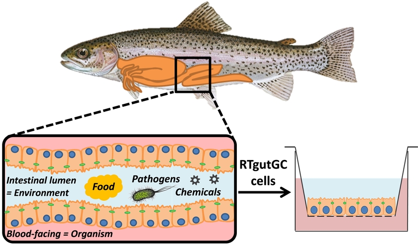
Das Darmepithel ist eine lebenswichtige Barriere. Es regelt die Aufnahme von Nahrung und bietet gleichermaßen Schutz vor Umweltgiften und Krankheitserreger. In unserer Gruppe haben wir ein in-vitro Model der Fischdarm-Barriere etabliert und charakterisiert. Es basiert auf der aus einer Regenbogenforelle (Onchorhynchus mykiss) isolierten Darmepithelzelllinie (RTgutGC). Dieses Modell wird verwendet, um die Auswirkungen von Umweltstressoren wie Nanopartikeln, Chemikalien und Immunstimulanzien systematisch zu untersuchen.
array(7 items)0 => Snowflake\Publications\Domain\Model\Publicationprototypepersistent entity (uid=19519, pid=124)originalId => protected19519 (integer)
authors => protected'Schug, H.; Maner, J.; Begnaud, F.; Berthaud, F.; Gimeno, S.; Schirmer, K.; Županič, A.' (123 chars)
title => protected'Intestinal fish cell barrier model to assess transfer of organic chemicals i n vitro: an experimental and computational study' (124 chars)
journal => protected'Environmental Science and Technology' (36 chars)
year => protected2019 (integer)
volume => protected53 (integer)
issue => protected'20' (2 chars)
startpage => protected'12062' (5 chars)
otherpage => protected'12070' (5 chars)
categories => protected'' (0 chars)
description => protected'We studied the role of the fish intestine as a barrier for organic chemicals using the epithelial barrier model built on the rainbow trout (<em>Oncorhyn chus mykiss</em>) intestinal cell line, RTgutGC and the newly developed expo sure chamber, TransFEr, specifically designed to work with hydrophobic and v olatile chemicals. Testing 11 chemicals with a range of physicochemical prop erties (logK<sub>OW</sub>: 2.2 to 6.3, logHLC: 6.1 to 2.3) and combining the data with a mechanistic kinetic model enabled the determination of dominant processes underlying the transfer experiments and the derivation of robust transfer rates. Against the current assumption in chemical uptake modeling, chemical transfer did not strictly depend on the logK<sub>OW</sub> but resul ted from chemical-specific intracellular accumulation and biotransformation combined with paracellular and active transport. Modeling also identified th at conducting elaborate measurements of the plastic parts, including the pol ystyrene insert and the PET filter, is unnecessary and that stirring in the TransFEr chamber reduced the stagnant water layers compared to theoretical p redictions. Aside from providing insights into chemical uptake via the intes tinal epithelium, this system can easily be transferred to other cell-based barrier systems, such as the fish gill or mammalian intestinal models and ma y improve in vitro–in vivo extrapolation and prediction of chemical bioacc umulation into organisms.' (1469 chars)
serialnumber => protected'0013-936X' (9 chars)
doi => protected'10.1021/acs.est.9b04281' (23 chars)
uid => protected19519 (integer)
_localizedUid => protected19519 (integer)modified_languageUid => protectedNULL
_versionedUid => protected19519 (integer)modifiedpid => protected124 (integer)1 => Snowflake\Publications\Domain\Model\Publicationprototypepersistent entity (uid=18485, pid=124)originalId => protected18485 (integer)
authors => protected'Schug, H.; Yue, Y.; Krese, R.; Fischer, S.; Kortner,&nbs p;T. M.; Schirmer, K.' (107 chars)
title => protected'Time- and concentration-dependent expression of immune and barrier genes in the RTgutGC fish intestinal model following immune stimulation' (138 chars)
journal => protected'Fish and Shellfish Immunology' (29 chars)
year => protected2019 (integer)
volume => protected88 (integer)
issue => protected'' (0 chars)
startpage => protected'308' (3 chars)
otherpage => protected'317' (3 chars)
categories => protected'rainbow trout; intestinal barrier; immune stimulation; LPS; Poly(I:C); cell culture model' (89 chars)
description => protected'The fish intestine comprises an important environment-organism interface tha t is vital to fish growth, health and pathogen defense. Yet, knowledge about the physiology and defense mechanisms toward environmental stressors, such as bacterial or viral cues, is limited and depends largely on <i>in vivo</i> experiments with fish. On this background, we here explore the immune compe tence of a recently established <i>in vitro</i> intestinal barrier model bas ed on the rainbow trout (<i>Oncorhynchus mykiss</i>) intestinal epithelial c ell line, RTgutGC. We demonstrate that the RTgutGC cell barrier reacts to tw o immune stimuli, the bacterial lipopolysaccharide (LPS) from <i>Escherichia coli</i> and the viral Poly(I:C), by regulating the mRNA abundance of selec ted genes in a partly time- and concentration dependent manner. The immune s timuli activated the Myd88-and Ticam-dependent signalling cascades, which re sulted in downstream activation of pro-inflammatory cytokines and interferon , comparable to the regulatory patterns known from <i>in vivo</i>. Stimuli e xposure furthermore influenced the regulation of epithelial barrier markers and resulted in slightly impaired barrier functionality after long-term expo sure to LPS. Collectively, we provide proof of the usefulness of this unique cell culture model to further gain basic understanding of the fish innate i mmune system and to apply it in various fields, such as fish feed developmen t and fish health in aquaculture or the evaluation of immuno-toxicity of che mical contaminants.' (1539 chars)
serialnumber => protected'1050-4648' (9 chars)
doi => protected'10.1016/j.fsi.2019.02.036' (25 chars)
uid => protected18485 (integer)
_localizedUid => protected18485 (integer)modified_languageUid => protectedNULL
_versionedUid => protected18485 (integer)modifiedpid => protected124 (integer)2 => Snowflake\Publications\Domain\Model\Publicationprototypepersistent entity (uid=18406, pid=124)originalId => protected18406 (integer)
authors => protected'Wang, J.; Lei, P.; Gamil, A. A. A.; Lagos, L.; Yue, Y.; Schirmer, K.; Mydland, L. T.; Øverland, M.; Krogdahl, Å.; Kortner, T. M.' (200 chars)
title => protected'Rainbow trout (<em>Oncorhynchus mykiss</em>) intestinal epithelial cells as a model for studying gut immune function and effects of functional feed ingr edients' (159 chars)
journal => protected'Frontiers in Immunology' (23 chars)
year => protected2019 (integer)
volume => protected10 (integer)
issue => protected'' (0 chars)
startpage => protected'152 (17 pp.)' (12 chars)
otherpage => protected'' (0 chars)
categories => protected'RTgutGC; in vitro model; lipopolysaccharide; functional feed ingredients; mu cosal immune responses; gut barrier' (111 chars)
description => protected'The objective of this study was to evaluate the suitability of the rainbow t rout intestinal epithelial cell line (RTgutGC) as an in vitro model for stud ies of gut immune function and effects of functional feed ingredients. Effec ts of lipopolysaccharide (LPS) and three functional feed ingredients [nucleo tides, mannanoligosaccharides (MOS), and beta-glucans] were evaluated in RTg utGC cells grown on conventional culture plates and transwell membranes. Per meation of fluorescently-labeled albumin, transepithelial electrical resista nce (TEER), and tight junction protein expression confirmed the barrier func tion of the cells. Brush border membrane enzyme activities [leucine aminopep tidase (LAP) and maltase] were detected in the RTgutGC cells but activity le vels were not modulated by any of the exposures. Immune related genes were e xpressed at comparable relative basal levels as these in rainbow trout dista l intestine. LPS produced markedly elevated gene expression levels of the pr oinflammatory cytokines <em>il1b</em>, <em>il6</em>, <em>il8</em>, and <em>t nfa</em> but had no effect on ROS production. Immunostaining demonstrated in creased F-actin contents after LPS exposure. Among the functional feed ingre dients, MOS seemed to be the most potent modulator of RTgutGC immune and bar rier function. MOS significantly increased albumin permeation and <em>il1b</ em>, <em>il6</em>, <em>il8</em>, <em>tnfa</em>, and <em>tgfb</em> expression , but suppressed ROS production, cell proliferation and <em>myd88</em> expre ssion. Induced levels of <em>il1b</em> and <em>il8</em> were also observed a fter treatment with nucleotides and beta-glucans. For barrier function relat ed genes, all treatments up-regulated the expression of <em>cldn3</em> and s uppressed <em>cdh1</em> levels. Beta-glucans increased TEER levels and F-act in content. Collectively, the present study has provided new information on how functional ingredients commonly applied in aquafeeds can affect intestin al epithelial function i...' (2320 chars)
serialnumber => protected'' (0 chars)
doi => protected'10.3389/fimmu.2019.00152' (24 chars)
uid => protected18406 (integer)
_localizedUid => protected18406 (integer)modified_languageUid => protectedNULL
_versionedUid => protected18406 (integer)modifiedpid => protected124 (integer)3 => Snowflake\Publications\Domain\Model\Publicationprototypepersistent entity (uid=17228, pid=124)originalId => protected17228 (integer)
authors => protected'Schug, H.; Begnaud, F.; Debonneville, C.; Berthaud, F.; Gimeno, S.; Schirmer, K.' (110 chars)
title => protected'TransFEr: a new device to measure the transfer of volatile and hydrophobic o rganic chemicals across an <i>in vitro</i> intestinal fish cell barrier' (147 chars)
journal => protected'Analytical Methods' (18 chars)
year => protected2018 (integer)
volume => protected10 (integer)
issue => protected'36' (2 chars)
startpage => protected'4394' (4 chars)
otherpage => protected'4403' (4 chars)
categories => protected'' (0 chars)
description => protected'Transfer of compounds across cellular barriers is a critical step of compoun d uptake into organisms. Using <i>in vitro</i> barrier systems to evaluate s uch transfer is attractive because of the higher throughput and reduced reso urce needs compared to animal studies. Thus far, however, studying the trans fer of hydrophobic and volatile compounds was hampered by the unavailability of <i>in vitro</i> exposure systems that allow for stable and predictable c hemical exposure concentrations. To overcome this limitation, we constructed a novel exposure chamber, TransFEr, and tested it with an <i>in vitro</i> e pithelial barrier model using the rainbow trout (<i>Oncorhynchus mykiss</i>) intestinal cell line, RTgutGC. Key features of the chamber are its closed d esign and rotatable silicon segments, which can serve for chemical dosing an d sampling. Using the fragrance damascone beta (log <i>K</i><sub>OW</sub>: 3.7, log HLC: −3.9) as a pilot chemical, we were able to demonstrate th at our exposure chamber provides for stable chemical exposure concentrations and full mass balance. The RTgutGC epithelium served as barrier for damasco ne beta transfer, which we attribute to chemical retention and biotransforma tion in the intestinal cells. Nevertheless, substantial transfer of the chem ical across the epithelium occurred. When a chemical sink, i.e. a silicon se gment, was included in the basolateral chamber to mimic blood constituents b inding <i>in vitro</i>, transfer was about three-fold enhanced. We suggest t hat the presented methodology can help to obtain insights into chemical upta ke mechanisms via the intestinal or other epithelia of fish and other animal s for hydrophobic and volatile chemicals.' (1713 chars)
serialnumber => protected'1759-9660' (9 chars)
doi => protected'10.1039/C8AY01253A' (18 chars)
uid => protected17228 (integer)
_localizedUid => protected17228 (integer)modified_languageUid => protectedNULL
_versionedUid => protected17228 (integer)modifiedpid => protected124 (integer)4 => Snowflake\Publications\Domain\Model\Publicationprototypepersistent entity (uid=14224, pid=124)originalId => protected14224 (integer)
authors => protected'Minghetti, M.; Drieschner, C.; Bramaz, N.; Schug, H.; Sc hirmer, K.' (91 chars)
title => protected'A fish intestinal epithelial barrier model established from the rainbow trou t (<I>Oncorhynchus mykiss</I>) cell line, RTgutGC' (125 chars)
journal => protected'Cell Biology and Toxicology' (27 chars)
year => protected2017 (integer)
volume => protected33 (integer)
issue => protected'6' (1 chars)
startpage => protected'539' (3 chars)
otherpage => protected'555' (3 chars)
categories => protected'fish intestine; polarized epithelium; permeation; ion regulation; silver tox icity; in vitro model' (97 chars)
description => protected'The intestine of fish is a multifunctional organ: lined by only a single lay er of specialized epithelial cells, it has various physiological roles inclu ding nutrient absorption and ion regulation. It moreover comprises an import ant barrier for environmental toxicants, including metals. Thus far, knowled ge of the fish intestine is limited largely to in vivo or ex vivo investigat ions. Recently, however, the first fish intestinal cell line, RTgutGC, was e stablished, originating from a rainbow trout (<I>Oncorhynchus mykiss</I>). I n order to exploit the opportunities arising from RTgutGC cells for explorin g fish intestinal physiology and toxicology, we present here the establishme nt of cells on commercially available permeable membrane supports and evalua te its suitability as a model of polarized intestinal epithelia. Within 3 we eks of culture, RTgutGC cells show epithelial features by forming tight junc tions and desmosomes between adjacent cells. Cells develop a transepithelial electrical resistance comparable to in vivo measured values, reflecting the leaky nature of the fish intestine. Immunocytochemistry reveals evidence of polarization, such as basolateral localization of Na<SUP>+</SUP>/K<SUP>+</S UP>-ATPase (NKA) and apical localization of the tight junction protein ZO-1. NKA mRNA abundance was induced as physiological response toward a saltwater buffer, mimicking the migration of rainbow trout from fresh to seawater. Pe rmeation of fluorescent molecules proved the barrier function of the cells, with permeation coefficients being comparable to those reported in fish. Fin ally, we demonstrate that cells on permeable supports are more resistant to the toxicity elicited by silver ions than cells grown the conventional way, likely due to improved cellular silver excretion.' (1797 chars)
serialnumber => protected'0742-2091' (9 chars)
doi => protected'10.1007/s10565-017-9385-x' (25 chars)
uid => protected14224 (integer)
_localizedUid => protected14224 (integer)modified_languageUid => protectedNULL
_versionedUid => protected14224 (integer)modifiedpid => protected124 (integer)5 => Snowflake\Publications\Domain\Model\Publicationprototypepersistent entity (uid=13949, pid=124)originalId => protected13949 (integer)
authors => protected'Drieschner, C.; Minghetti, M.; Wu, S.; Renaud, P.; Schir mer, K.' (88 chars)
title => protected'Ultrathin alumina membranes as scaffold for epithelial cell culture from the intestine of rainbow trout' (103 chars)
journal => protected'ACS Applied Materials and Interfaces' (36 chars)
year => protected2017 (integer)
volume => protected9 (integer)
issue => protected'11' (2 chars)
startpage => protected'9496' (4 chars)
otherpage => protected'9505' (4 chars)
categories => protected'fish-gut-on-chip; in vitro epithelial barrier model; ultrathin membrane; ano dized aluminum; cell culture interface; impedance based toxicity testing' (148 chars)
description => protected'Permeable membranes are indispensable for in vitro epithelial barrier models . However, currently available polymer-based membranes are low in porosity a nd relatively thick, resulting in a limited permeability and unrealistic cul ture conditions. In this study, we developed an ultrathin, nanoporous alumin a membrane as novel cell culture interface for vertebrate cells, with focus on the rainbow trout (<I>Onchorynchus mykiss</I>) intestinal cell line RTgut GC. The new type of membrane is framed in a silicon chip for physical suppor t and has a thickness of only 1 µm, with a porosity of 15% and homogeneous nanopores (Ø = 73 ± 21 nm). Permeability rates for small molecules, namely lucifer yellow, dextran 40 and bovine serum albumin, exceeded those of stan dard polyethylene terephthalate (PET) membranes by up to 27 fold. With the f inal goal to establish a representative model of the fish intestine for envi ronmental toxicology, we engineered a simple culture set-up, capable to test the cellular response towards chemical exposure. Herein, cells were culture d in a monolayer on the alumina membranes and formed a polarized epithelium with apical expression of the tight junction protein ZO-1 within 14 days. Im pedance spectroscopy, a non-invasive and real time electrical measurement, w as used to determine cellular resistance during epithelial layer formation a nd chemical exposure to evaluate barrier functionality. Resistance values du ring epithelial development revealed different stages of epithelial maturity and were comparable with the in vivo situation. During chemical exposure, c ellular resistance changed immediately, when barrier tightness or cell viabi lity was affected. Thus, our study demonstrates nanoporous alumina membranes as promising novel interface for alterative in vitro approaches, capable to allow cell culture in a physiologically realistic manner and to enable high quality microscopy and sensitive measurement of cellular resistance.' (1969 chars)
serialnumber => protected'1944-8244' (9 chars)
doi => protected'10.1021/acsami.7b00705' (22 chars)
uid => protected13949 (integer)
_localizedUid => protected13949 (integer)modified_languageUid => protectedNULL
_versionedUid => protected13949 (integer)modifiedpid => protected124 (integer)6 => Snowflake\Publications\Domain\Model\Publicationprototypepersistent entity (uid=10589, pid=124)originalId => protected10589 (integer)
authors => protected'Geppert, M.; Sigg, L.; Schirmer, K.' (50 chars)
title => protected'A novel two-compartment barrier model for investigating nanoparticle transpo rt in fish intestinal epithelial cells' (114 chars)
journal => protected'Environmental Science: Nano' (27 chars)
year => protected2016 (integer)
volume => protected3 (integer)
issue => protected'2' (1 chars)
startpage => protected'388' (3 chars)
otherpage => protected'395' (3 chars)
categories => protected'' (0 chars)
description => protected'We introduce a novel <em>in vitro</em> rainbow trout intestinal barrier mode l and demonstrate its suitability for investigating nanoparticle transport a cross the intestinal epithelium. Rainbow trout (<em>Oncorhynchus mykiss</em> ) intestinal cells (RTgutGC) were grown as monolayers on permeable supports leading to a two-compartment intestinal barrier model consisting of a polari zed epithelium, dividing the system into an upper (apical) and a lower (baso lateral) compartment, and thereby mimicking the intestinal lumen and the por tal blood, respectively. The cells express the tight junction protein ZO-1 a nd build up a transepithelial electrical resistance comparable to the <em>in vivo</em> situation. Fluorescent polystyrene nanoparticles (PS-NPs; average hydrodynamic diameter: 73 ± 18 nm) were accumulated by RTgutGC cells in a time-, temperature- and concentration-dependent manner. Uptake of PS-NPs was confirmed using fluorescence microscopy. Cells formed an efficient barrier largely preventing the translocation of PS-NPs to the basolateral compartmen t. Taken together, these data demonstrate the suitability of the <em>in vitr o</em> barrier model to study the effects of nanoparticles in fish intestina l epithelial cells.' (1235 chars)
serialnumber => protected'2051-8153' (9 chars)
doi => protected'10.1039/c5en00226e' (18 chars)
uid => protected10589 (integer)
_localizedUid => protected10589 (integer)modified_languageUid => protectedNULL
_versionedUid => protected10589 (integer)modifiedpid => protected124 (integer)
Intestinal fish cell barrier model to assess transfer of organic chemicals in vitro: an experimental and computational study
We studied the role of the fish intestine as a barrier for organic chemicals using the epithelial barrier model built on the rainbow trout (Oncorhynchus mykiss) intestinal cell line, RTgutGC and the newly developed exposure chamber, TransFEr, specifically designed to work with hydrophobic and volatile chemicals. Testing 11 chemicals with a range of physicochemical properties (logKOW: 2.2 to 6.3, logHLC: 6.1 to 2.3) and combining the data with a mechanistic kinetic model enabled the determination of dominant processes underlying the transfer experiments and the derivation of robust transfer rates. Against the current assumption in chemical uptake modeling, chemical transfer did not strictly depend on the logKOW but resulted from chemical-specific intracellular accumulation and biotransformation combined with paracellular and active transport. Modeling also identified that conducting elaborate measurements of the plastic parts, including the polystyrene insert and the PET filter, is unnecessary and that stirring in the TransFEr chamber reduced the stagnant water layers compared to theoretical predictions. Aside from providing insights into chemical uptake via the intestinal epithelium, this system can easily be transferred to other cell-based barrier systems, such as the fish gill or mammalian intestinal models and may improve in vitro–in vivo extrapolation and prediction of chemical bioaccumulation into organisms.
Schug, H.; Maner, J.; Begnaud, F.; Berthaud, F.; Gimeno, S.; Schirmer, K.; Županič, A. (2019) Intestinal fish cell barrier model to assess transfer of organic chemicals in vitro: an experimental and computational study, Environmental Science and Technology, 53(20), 12062-12070, doi:10.1021/acs.est.9b04281, Institutional Repository
Time- and concentration-dependent expression of immune and barrier genes in the RTgutGC fish intestinal model following immune stimulation
The fish intestine comprises an important environment-organism interface that is vital to fish growth, health and pathogen defense. Yet, knowledge about the physiology and defense mechanisms toward environmental stressors, such as bacterial or viral cues, is limited and depends largely on in vivo experiments with fish. On this background, we here explore the immune competence of a recently established in vitro intestinal barrier model based on the rainbow trout (Oncorhynchus mykiss) intestinal epithelial cell line, RTgutGC. We demonstrate that the RTgutGC cell barrier reacts to two immune stimuli, the bacterial lipopolysaccharide (LPS) from Escherichia coli and the viral Poly(I:C), by regulating the mRNA abundance of selected genes in a partly time- and concentration dependent manner. The immune stimuli activated the Myd88-and Ticam-dependent signalling cascades, which resulted in downstream activation of pro-inflammatory cytokines and interferon, comparable to the regulatory patterns known from in vivo. Stimuli exposure furthermore influenced the regulation of epithelial barrier markers and resulted in slightly impaired barrier functionality after long-term exposure to LPS. Collectively, we provide proof of the usefulness of this unique cell culture model to further gain basic understanding of the fish innate immune system and to apply it in various fields, such as fish feed development and fish health in aquaculture or the evaluation of immuno-toxicity of chemical contaminants.
Schug, H.; Yue, Y.; Krese, R.; Fischer, S.; Kortner, T. M.; Schirmer, K. (2019) Time- and concentration-dependent expression of immune and barrier genes in the RTgutGC fish intestinal model following immune stimulation, Fish and Shellfish Immunology, 88, 308-317, doi:10.1016/j.fsi.2019.02.036, Institutional Repository
Rainbow trout (Oncorhynchus mykiss) intestinal epithelial cells as a model for studying gut immune function and effects of functional feed ingredients
The objective of this study was to evaluate the suitability of the rainbow trout intestinal epithelial cell line (RTgutGC) as an in vitro model for studies of gut immune function and effects of functional feed ingredients. Effects of lipopolysaccharide (LPS) and three functional feed ingredients [nucleotides, mannanoligosaccharides (MOS), and beta-glucans] were evaluated in RTgutGC cells grown on conventional culture plates and transwell membranes. Permeation of fluorescently-labeled albumin, transepithelial electrical resistance (TEER), and tight junction protein expression confirmed the barrier function of the cells. Brush border membrane enzyme activities [leucine aminopeptidase (LAP) and maltase] were detected in the RTgutGC cells but activity levels were not modulated by any of the exposures. Immune related genes were expressed at comparable relative basal levels as these in rainbow trout distal intestine. LPS produced markedly elevated gene expression levels of the proinflammatory cytokines il1b, il6, il8, and tnfa but had no effect on ROS production. Immunostaining demonstrated increased F-actin contents after LPS exposure. Among the functional feed ingredients, MOS seemed to be the most potent modulator of RTgutGC immune and barrier function. MOS significantly increased albumin permeation and il1b, il6, il8, tnfa, and tgfb expression, but suppressed ROS production, cell proliferation and myd88 expression. Induced levels of il1b and il8 were also observed after treatment with nucleotides and beta-glucans. For barrier function related genes, all treatments up-regulated the expression of cldn3 and suppressed cdh1 levels. Beta-glucans increased TEER levels and F-actin content. Collectively, the present study has provided new information on how functional ingredients commonly applied in aquafeeds can affect intestinal epithelial function in fish. Our findings suggest that RTgutGC cells possess characteristic features of functional intestinal epithelial cells indicating a potential for use as an efficient in vitro model to evaluate effects of bioactive feed ingredients on gut immune and barrier functions and their underlying cellular mechanisms.
Wang, J.; Lei, P.; Gamil, A. A. A.; Lagos, L.; Yue, Y.; Schirmer, K.; Mydland, L. T.; Øverland, M.; Krogdahl, Å.; Kortner, T. M. (2019) Rainbow trout (Oncorhynchus mykiss) intestinal epithelial cells as a model for studying gut immune function and effects of functional feed ingredients, Frontiers in Immunology, 10, 152 (17 pp.), doi:10.3389/fimmu.2019.00152, Institutional Repository
TransFEr: a new device to measure the transfer of volatile and hydrophobic organic chemicals across an in vitro intestinal fish cell barrier
Transfer of compounds across cellular barriers is a critical step of compound uptake into organisms. Using in vitro barrier systems to evaluate such transfer is attractive because of the higher throughput and reduced resource needs compared to animal studies. Thus far, however, studying the transfer of hydrophobic and volatile compounds was hampered by the unavailability of in vitro exposure systems that allow for stable and predictable chemical exposure concentrations. To overcome this limitation, we constructed a novel exposure chamber, TransFEr, and tested it with an in vitro epithelial barrier model using the rainbow trout (Oncorhynchus mykiss) intestinal cell line, RTgutGC. Key features of the chamber are its closed design and rotatable silicon segments, which can serve for chemical dosing and sampling. Using the fragrance damascone beta (log KOW: 3.7, log HLC: −3.9) as a pilot chemical, we were able to demonstrate that our exposure chamber provides for stable chemical exposure concentrations and full mass balance. The RTgutGC epithelium served as barrier for damascone beta transfer, which we attribute to chemical retention and biotransformation in the intestinal cells. Nevertheless, substantial transfer of the chemical across the epithelium occurred. When a chemical sink, i.e. a silicon segment, was included in the basolateral chamber to mimic blood constituents binding in vitro, transfer was about three-fold enhanced. We suggest that the presented methodology can help to obtain insights into chemical uptake mechanisms via the intestinal or other epithelia of fish and other animals for hydrophobic and volatile chemicals.
Schug, H.; Begnaud, F.; Debonneville, C.; Berthaud, F.; Gimeno, S.; Schirmer, K. (2018) TransFEr: a new device to measure the transfer of volatile and hydrophobic organic chemicals across an in vitro intestinal fish cell barrier, Analytical Methods, 10(36), 4394-4403, doi:10.1039/C8AY01253A, Institutional Repository
A fish intestinal epithelial barrier model established from the rainbow trout (Oncorhynchus mykiss) cell line, RTgutGC
The intestine of fish is a multifunctional organ: lined by only a single layer of specialized epithelial cells, it has various physiological roles including nutrient absorption and ion regulation. It moreover comprises an important barrier for environmental toxicants, including metals. Thus far, knowledge of the fish intestine is limited largely to in vivo or ex vivo investigations. Recently, however, the first fish intestinal cell line, RTgutGC, was established, originating from a rainbow trout (Oncorhynchus mykiss). In order to exploit the opportunities arising from RTgutGC cells for exploring fish intestinal physiology and toxicology, we present here the establishment of cells on commercially available permeable membrane supports and evaluate its suitability as a model of polarized intestinal epithelia. Within 3 weeks of culture, RTgutGC cells show epithelial features by forming tight junctions and desmosomes between adjacent cells. Cells develop a transepithelial electrical resistance comparable to in vivo measured values, reflecting the leaky nature of the fish intestine. Immunocytochemistry reveals evidence of polarization, such as basolateral localization of Na+/K+-ATPase (NKA) and apical localization of the tight junction protein ZO-1. NKA mRNA abundance was induced as physiological response toward a saltwater buffer, mimicking the migration of rainbow trout from fresh to seawater. Permeation of fluorescent molecules proved the barrier function of the cells, with permeation coefficients being comparable to those reported in fish. Finally, we demonstrate that cells on permeable supports are more resistant to the toxicity elicited by silver ions than cells grown the conventional way, likely due to improved cellular silver excretion.
Minghetti, M.; Drieschner, C.; Bramaz, N.; Schug, H.; Schirmer, K. (2017) A fish intestinal epithelial barrier model established from the rainbow trout (Oncorhynchus mykiss) cell line, RTgutGC, Cell Biology and Toxicology, 33(6), 539-555, doi:10.1007/s10565-017-9385-x, Institutional Repository
Ultrathin alumina membranes as scaffold for epithelial cell culture from the intestine of rainbow trout
Permeable membranes are indispensable for in vitro epithelial barrier models. However, currently available polymer-based membranes are low in porosity and relatively thick, resulting in a limited permeability and unrealistic culture conditions. In this study, we developed an ultrathin, nanoporous alumina membrane as novel cell culture interface for vertebrate cells, with focus on the rainbow trout (Onchorynchus mykiss) intestinal cell line RTgutGC. The new type of membrane is framed in a silicon chip for physical support and has a thickness of only 1 µm, with a porosity of 15% and homogeneous nanopores (Ø = 73 ± 21 nm). Permeability rates for small molecules, namely lucifer yellow, dextran 40 and bovine serum albumin, exceeded those of standard polyethylene terephthalate (PET) membranes by up to 27 fold. With the final goal to establish a representative model of the fish intestine for environmental toxicology, we engineered a simple culture set-up, capable to test the cellular response towards chemical exposure. Herein, cells were cultured in a monolayer on the alumina membranes and formed a polarized epithelium with apical expression of the tight junction protein ZO-1 within 14 days. Impedance spectroscopy, a non-invasive and real time electrical measurement, was used to determine cellular resistance during epithelial layer formation and chemical exposure to evaluate barrier functionality. Resistance values during epithelial development revealed different stages of epithelial maturity and were comparable with the in vivo situation. During chemical exposure, cellular resistance changed immediately, when barrier tightness or cell viability was affected. Thus, our study demonstrates nanoporous alumina membranes as promising novel interface for alterative in vitro approaches, capable to allow cell culture in a physiologically realistic manner and to enable high quality microscopy and sensitive measurement of cellular resistance.
Drieschner, C.; Minghetti, M.; Wu, S.; Renaud, P.; Schirmer, K. (2017) Ultrathin alumina membranes as scaffold for epithelial cell culture from the intestine of rainbow trout, ACS Applied Materials and Interfaces, 9(11), 9496-9505, doi:10.1021/acsami.7b00705, Institutional Repository
A novel two-compartment barrier model for investigating nanoparticle transport in fish intestinal epithelial cells
We introduce a novel in vitro rainbow trout intestinal barrier model and demonstrate its suitability for investigating nanoparticle transport across the intestinal epithelium. Rainbow trout (Oncorhynchus mykiss) intestinal cells (RTgutGC) were grown as monolayers on permeable supports leading to a two-compartment intestinal barrier model consisting of a polarized epithelium, dividing the system into an upper (apical) and a lower (basolateral) compartment, and thereby mimicking the intestinal lumen and the portal blood, respectively. The cells express the tight junction protein ZO-1 and build up a transepithelial electrical resistance comparable to the in vivo situation. Fluorescent polystyrene nanoparticles (PS-NPs; average hydrodynamic diameter: 73 ± 18 nm) were accumulated by RTgutGC cells in a time-, temperature- and concentration-dependent manner. Uptake of PS-NPs was confirmed using fluorescence microscopy. Cells formed an efficient barrier largely preventing the translocation of PS-NPs to the basolateral compartment. Taken together, these data demonstrate the suitability of the in vitro barrier model to study the effects of nanoparticles in fish intestinal epithelial cells.
Geppert, M.; Sigg, L.; Schirmer, K. (2016) A novel two-compartment barrier model for investigating nanoparticle transport in fish intestinal epithelial cells, Environmental Science: Nano, 3(2), 388-395, doi:10.1039/c5en00226e, Institutional Repository