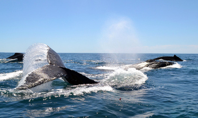
In Zusammenarbeit mit der Griffith University (Brisbane) haben wir Buckelwal-Zelllinien entwickelt. HuWawild-type stammen aus Hautproben von freilebenden Individuen und HuWaTERT wurden mit TERT transfiziert, was eine langfristige Kultivierung und Anwendung sicherstellt.
Die Zelllinien werden in der Ökotoxikologie verwendet, um die Auswirkungen von relevanten Chemikalien in vitro zu untersuchen und ermöglichen dadurch eine schnelle und kostengünstige Toxizitätsbewertung.
array(3 items)0 => Snowflake\Publications\Domain\Model\Publicationprototypepersistent entity (uid=19798, pid=124)originalId => protected19798 (integer)
authors => protected'Maner, J.; Burkard, M.; Cassano, J. C.; Bengtson Nash,&n bsp;S. M.; Schirmer, K.; Suter, M. J. -F.' (142 chars)
title => protected'Hexachlorobenzene exerts genotoxic effects in a humpback whale cell line und er stable exposure conditions' (105 chars)
journal => protected'RSC Advances' (12 chars)
year => protected2019 (integer)
volume => protected9 (integer)
issue => protected'67' (2 chars)
startpage => protected'39447' (5 chars)
otherpage => protected'39457' (5 chars)
categories => protected'' (0 chars)
description => protected'Humpback whales, like other polar wildlife, accumulate persistent organic po llutants. In Southern hemisphere populations, hexachlorobenzene (HCB) domina tes the contaminant profiles. HCB is linked to a variety of health effects a nd is classified as a group 2B carcinogen, but the mechanism of action is a matter of contention. Potential toxicological effects to humpback whales rem ain entirely unknown. The recently established humpback whale fibroblast cel l line (HuWa) offers an <em>in vitro</em> model for toxicological investigat ions. We here combine this novel cell line with a passive dosing strategy to investigate whale-specific toxicity of HCB. The relevant partitioning coeff icients were determined to produce stable and predictable exposure concentra tions in small-scale bioassays. The system was used to assess acute toxicity as well as genotoxicity of HCB to the HuWa cell line. While we found some t ransient reductions in metabolic activity, measured with the indicator dye a lamarBlue, no clear acute toxic effects were discernible. Yet, a significant increase in DNA damage, detected in the alkaline comet assay, was found in HuWa cells exposed to 10 μg L<sup>-1</sup> HCB during the sensitive phase o f cell attachment. Collectively, this work provides a ready-to-use passive d osing system and delivers evidence that HCB elicits genotoxicity in humpback whale cells.' (1381 chars)
serialnumber => protected'2046-2069' (9 chars)
doi => protected'10.1039/C9RA05352B' (18 chars)
uid => protected19798 (integer)
_localizedUid => protected19798 (integer)modified_languageUid => protectedNULL
_versionedUid => protected19798 (integer)modifiedpid => protected124 (integer)1 => Snowflake\Publications\Domain\Model\Publicationprototypepersistent entity (uid=18228, pid=124)originalId => protected18228 (integer)
authors => protected'Burkard, M.; Bengtson Nash, S.; Gambaro, G.; Whitworth, D.; Schirmer, K.' (97 chars)
title => protected'Lifetime extension of humpback whale skin fibroblasts and their response to lipopolysaccharide (LPS) and a mixture of polychlorinated biphenyls (Aroclor )' (153 chars)
journal => protected'Cell Biology and Toxicology' (27 chars)
year => protected2019 (integer)
volume => protected35 (integer)
issue => protected'4' (1 chars)
startpage => protected'387' (3 chars)
otherpage => protected'398' (3 chars)
categories => protected'cell line transfection; humpback whale; Megaptera novaeangliae; immunotoxici ty; inflammatory cytokines; relative telomerase activity' (132 chars)
description => protected'Marine mammals, such as whales, have a high proportion of body fat and so ar e susceptible to the accumulation, and associated detrimental health effects , of lipophilic environmental contaminants. Recently, we developed a wild-ty pe cell line from humpback whale fibroblasts (HuWa). Extensive molecular ass essments with mammalian wild-type cells are typically constrained by a finit e life span, with cells eventually becoming senescent. Thus, the present wor k explored the possibility of preventing senescence in the HuWa cell line by transfection with plasmids encoding the simian virus large T antigen (SV40T ) or telomerase reverse transcriptase (TERT). No stable expression was achie ved upon SV40 transfection. Transfection with TERT, on the other hand, activ ated the expression of telomerase in HuWa cells. At the time of manuscript p reparation, the transfected HuWa cells (HuWa<sub>TERT</sub>) have been stabl e for at least 59 passages post-transfection. HuWa<sub>TERT</sub> proliferat e rapidly and maintain initial cell characteristics, such as morphology and chromosomal stability. The response of HuWa<sub>TERT</sub> cells to an immun e stimulant (lipopolysaccharide (LPS)) and an immunotoxicant (Aroclor1254) w as assessed by measurement of intracellular levels of the pro-inflammatory c ytokines interleukin (IL)-6, IL-1β and tumour necrosis factor (TNF)-α. HuW a<sub>TERT</sub> cells constitutively express IL-6, IL-1β and TNFα. Exposu re to neither LPS nor Aroclor1254 had an effect on the levels of these cytok ines. Overall, this work supports the diverse applicability of HuWa cell lin es in that they display reliable long-term preservation, susceptibility to e xogenous gene transfer and enable the study of humpback whale-specific cellu lar response mechanisms.' (1772 chars)
serialnumber => protected'0742-2091' (9 chars)
doi => protected'10.1007/s10565-018-09457-1' (26 chars)
uid => protected18228 (integer)
_localizedUid => protected18228 (integer)modified_languageUid => protectedNULL
_versionedUid => protected18228 (integer)modifiedpid => protected124 (integer)2 => Snowflake\Publications\Domain\Model\Publicationprototypepersistent entity (uid=8262, pid=124)originalId => protected8262 (integer)
authors => protected'Burkard, M.; Whitworth, D.; Schirmer, K.; Nash, S. B.' (78 chars)
title => protected'Establishment of the first humpback whale fibroblast cell lines and their ap plication in chemical risk assessment' (113 chars)
journal => protected'Aquatic Toxicology' (18 chars)
year => protected2015 (integer)
volume => protected167 (integer)
issue => protected'' (0 chars)
startpage => protected'240' (3 chars)
otherpage => protected'247' (3 chars)
categories => protected'Megaptera novaeangliae; p,p′,-DDE; Antarctica; persistent organic pollutan ts (POPs); karyotype; cell line characterization' (124 chars)
description => protected'This paper reports the first successful derivation and characterization of h umpback whale fibroblast cell lines. Primary fibroblasts were isolated from the dermal connective tissue of skin biopsies, cultured at 37 °C and 5% CO <SUB>2</SUB> in the standard mammalian medium DMEM/F12 supplemented with 10% fetal bovine serum (FBS). Of nine initial biopsies, two cell lines were est ablished from two different animals and designated HuWa1 and HuWa2. The cell s have a stable karyotype with 2<I>n</I> = 44, which has commonly been obs erved in other baleen whale species. Cells were verified as being fibroblast s based on their spindle-shaped morphology, adherence to plastic and positiv e immunoreaction to vimentin. Population doubling time was determined to be ∼41 h and cells were successfully cryopreserved and thawed. To date, HuWa 1 cells have been propagated 30 times. Cells proliferate at the tested tempe ratures, 30, 33.5 and 37 °C, but show the highest rate of proliferation at 37 °C. Short-term exposure to <I>para</I>,<I>para′</I>-dichlorodiphenyl dichloroethylene (<I>p,p′-</I>DDE), a priority compound accumulating in so uthern hemisphere humpback whales, resulted in a concentration-dependent los s of cell viability. The effective concentration which caused a 50% reductio n in HuWa1 cell viability (EC<SUB>50</SUB> value) was approximately six time s greater than the EC<SUB>50</SUB> value for the same chemical measured with human dermal fibroblasts. HuWa1 exposed to a natural, <I>p,p′</I>-DDE-con taining, chemical mixture extracted from whale blubber showed distinctively higher sensitivity than to <I>p,p′-</I>DDE alone. Thus, we provide the fir st cytotoxicological data for humpback whales and with establishment of the HuWa cell lines, a unique <I>in vitro</I> model for the study of the whales' sensitivity and cellular response to chemicals and other environmental stre ssors.' (1906 chars)
serialnumber => protected'0166-445X' (9 chars)
doi => protected'10.1016/j.aquatox.2015.08.005' (29 chars)
uid => protected8262 (integer)
_localizedUid => protected8262 (integer)modified_languageUid => protectedNULL
_versionedUid => protected8262 (integer)modifiedpid => protected124 (integer)
Hexachlorobenzene exerts genotoxic effects in a humpback whale cell line under stable exposure conditions
Humpback whales, like other polar wildlife, accumulate persistent organic pollutants. In Southern hemisphere populations, hexachlorobenzene (HCB) dominates the contaminant profiles. HCB is linked to a variety of health effects and is classified as a group 2B carcinogen, but the mechanism of action is a matter of contention. Potential toxicological effects to humpback whales remain entirely unknown. The recently established humpback whale fibroblast cell line (HuWa) offers an in vitro model for toxicological investigations. We here combine this novel cell line with a passive dosing strategy to investigate whale-specific toxicity of HCB. The relevant partitioning coefficients were determined to produce stable and predictable exposure concentrations in small-scale bioassays. The system was used to assess acute toxicity as well as genotoxicity of HCB to the HuWa cell line. While we found some transient reductions in metabolic activity, measured with the indicator dye alamarBlue, no clear acute toxic effects were discernible. Yet, a significant increase in DNA damage, detected in the alkaline comet assay, was found in HuWa cells exposed to 10 μg L-1 HCB during the sensitive phase of cell attachment. Collectively, this work provides a ready-to-use passive dosing system and delivers evidence that HCB elicits genotoxicity in humpback whale cells.
Maner, J.; Burkard, M.; Cassano, J. C.; Bengtson Nash, S. M.; Schirmer, K.; Suter, M. J. -F. (2019) Hexachlorobenzene exerts genotoxic effects in a humpback whale cell line under stable exposure conditions, RSC Advances, 9(67), 39447-39457, doi:10.1039/C9RA05352B, Institutional Repository
Lifetime extension of humpback whale skin fibroblasts and their response to lipopolysaccharide (LPS) and a mixture of polychlorinated biphenyls (Aroclor)
Marine mammals, such as whales, have a high proportion of body fat and so are susceptible to the accumulation, and associated detrimental health effects, of lipophilic environmental contaminants. Recently, we developed a wild-type cell line from humpback whale fibroblasts (HuWa). Extensive molecular assessments with mammalian wild-type cells are typically constrained by a finite life span, with cells eventually becoming senescent. Thus, the present work explored the possibility of preventing senescence in the HuWa cell line by transfection with plasmids encoding the simian virus large T antigen (SV40T) or telomerase reverse transcriptase (TERT). No stable expression was achieved upon SV40 transfection. Transfection with TERT, on the other hand, activated the expression of telomerase in HuWa cells. At the time of manuscript preparation, the transfected HuWa cells (HuWaTERT) have been stable for at least 59 passages post-transfection. HuWaTERT proliferate rapidly and maintain initial cell characteristics, such as morphology and chromosomal stability. The response of HuWaTERT cells to an immune stimulant (lipopolysaccharide (LPS)) and an immunotoxicant (Aroclor1254) was assessed by measurement of intracellular levels of the pro-inflammatory cytokines interleukin (IL)-6, IL-1β and tumour necrosis factor (TNF)-α. HuWaTERT cells constitutively express IL-6, IL-1β and TNFα. Exposure to neither LPS nor Aroclor1254 had an effect on the levels of these cytokines. Overall, this work supports the diverse applicability of HuWa cell lines in that they display reliable long-term preservation, susceptibility to exogenous gene transfer and enable the study of humpback whale-specific cellular response mechanisms.
Burkard, M.; Bengtson Nash, S.; Gambaro, G.; Whitworth, D.; Schirmer, K. (2019) Lifetime extension of humpback whale skin fibroblasts and their response to lipopolysaccharide (LPS) and a mixture of polychlorinated biphenyls (Aroclor), Cell Biology and Toxicology, 35(4), 387-398, doi:10.1007/s10565-018-09457-1, Institutional Repository
Establishment of the first humpback whale fibroblast cell lines and their application in chemical risk assessment
This paper reports the first successful derivation and characterization of humpback whale fibroblast cell lines. Primary fibroblasts were isolated from the dermal connective tissue of skin biopsies, cultured at 37 °C and 5% CO2 in the standard mammalian medium DMEM/F12 supplemented with 10% fetal bovine serum (FBS). Of nine initial biopsies, two cell lines were established from two different animals and designated HuWa1 and HuWa2. The cells have a stable karyotype with 2n = 44, which has commonly been observed in other baleen whale species. Cells were verified as being fibroblasts based on their spindle-shaped morphology, adherence to plastic and positive immunoreaction to vimentin. Population doubling time was determined to be ∼41 h and cells were successfully cryopreserved and thawed. To date, HuWa1 cells have been propagated 30 times. Cells proliferate at the tested temperatures, 30, 33.5 and 37 °C, but show the highest rate of proliferation at 37 °C. Short-term exposure to para,para′-dichlorodiphenyldichloroethylene (p,p′-DDE), a priority compound accumulating in southern hemisphere humpback whales, resulted in a concentration-dependent loss of cell viability. The effective concentration which caused a 50% reduction in HuWa1 cell viability (EC50 value) was approximately six times greater than the EC50 value for the same chemical measured with human dermal fibroblasts. HuWa1 exposed to a natural, p,p′-DDE-containing, chemical mixture extracted from whale blubber showed distinctively higher sensitivity than to p,p′-DDE alone. Thus, we provide the first cytotoxicological data for humpback whales and with establishment of the HuWa cell lines, a unique in vitro model for the study of the whales' sensitivity and cellular response to chemicals and other environmental stressors.
Burkard, M.; Whitworth, D.; Schirmer, K.; Nash, S. B. (2015) Establishment of the first humpback whale fibroblast cell lines and their application in chemical risk assessment, Aquatic Toxicology, 167, 240-247, doi:10.1016/j.aquatox.2015.08.005, Institutional Repository