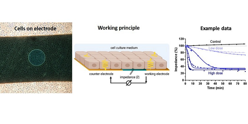
Microscopic image of cells on chip by J. Maner, working principle image created with BioRender, example data image by C. Drieschner
In the RAINBOflow Chip project, we are developing a biosensor system to detect toxic effects of chemicals by impedance sensing. At the core of the biosensor are cell lines from the rainbow trout (Oncorhynchus mykiss), which can predict toxic effects to fish. They are seeded on a microfluidic chip where their adherence to the electrodes creates a resistance to an applied electric current flow. This resistance reflects the health status of the cells; a decrease in resistance is an indicator for loss of cell viability. This method is quick, non-invasive, and can be automated, thus it allows monitoring toxicity in real-time. Our aim is to employ this method for time-resolved toxicity testing of chemicals under flow conditions, as well as to establish a portable and compact system for use in the field, with the data being accessible online to inform about the current water quality.
The portable biosensor installed on the LéXPLORE platform on lake Geneva for field testing (Photo: J. Maner)
array(5 items)0 => Snowflake\Publications\Domain\Model\Publicationprototypepersistent entity (uid=19144, pid=124)originalId => protected19144 (integer)
authors => protected'Drieschner, C.; Könemann, S.; Renaud, P.; Schirmer, K.' (75 chars)
title => protected'Fish-gut-on-chip: development of a microfluidic bioreactor to study the role of the fish intestine <em>in vitro</em>' (116 chars)
journal => protected'Lab on a Chip' (13 chars)
year => protected2019 (integer)
volume => protected19 (integer)
issue => protected'19' (2 chars)
startpage => protected'3268' (4 chars)
otherpage => protected'3276' (4 chars)
categories => protected'' (0 chars)
description => protected'In this study we present the first fish-gut-on-chip model. This model is bas ed on the reconstruction of the intestinal barrier by culturing two intestin al cell lines from rainbow trout, namely epithelial RTgutGC and fibroblastic RTgutF, in an artificial microenvironment. For a realistic mimicry of the i nterface between the intestinal lumen and the interior of the organism we i) developed ultrathin and highly porous silicon nitride membranes that serve as basement membrane analogues and provide a culture interface for the fish cells; ii) constructed a unique micro-well plate-based microfluidic bioreact or that enables parallelization of experiments and creates realistic fluid f low exposure scenarios for the cells; iii) integrated electrodes in the reac tor for non-invasive impedance sensing of cellular well-being. In a first ap proach, we used this reactor to investigate the response of epithelial fish cells to <em>in vivo</em>-like shear stress rates of 0.002–0.06 dyne per c m<sup>2</sup>, resulting from fluid flow within the intestinal lumen. Moreov er, we investigated the interplay of epithelial and fibroblast cells under o ptimal flow conditions to carefully evaluate the benefits and drawbacks of t he more complex reconstruction of the intestinal architecture. With our fish -gut-on-chip model we open up new strategies for a better understanding of b asic fish physiology, for the refinement of fish feed in aquaculture and for predicting chemical uptake and bioaccumulation in fish for environmental ri sk assessment. The basic principles of our reactor prototype, including the use of ultrathin membranes, an open microfluidic circuit for perfusion and t he micro-well plate-based format for simplified handling and avoidance of ai r-bubbles, will as well be of great value for other barrier-on-chip models.' (1823 chars)
serialnumber => protected'1473-0197' (9 chars)
doi => protected'10.1039/C9LC00415G' (18 chars)
uid => protected19144 (integer)
_localizedUid => protected19144 (integer)modified_languageUid => protectedNULL
_versionedUid => protected19144 (integer)modifiedpid => protected124 (integer)1 => Snowflake\Publications\Domain\Model\Publicationprototypepersistent entity (uid=18979, pid=124)originalId => protected18979 (integer)
authors => protected'Drieschner, C.; Vo, N. T. K.; Schug, H.; Burkard,&n bsp;M.; Bols, N. C.; Renaud, P.; Schirmer, K.' (141 chars)
title => protected'Improving a fish intestinal barrier model by combining two rainbow trout cel l lines: epithelial RTgutGC and fibroblastic RTgutF' (127 chars)
journal => protected'Cytotechnology' (14 chars)
year => protected2019 (integer)
volume => protected71 (integer)
issue => protected'4' (1 chars)
startpage => protected'835' (3 chars)
otherpage => protected'848' (3 chars)
categories => protected'fish-gut-on-chip; rainbow trout (Oncorhynchus mykiss); epithelial barrier mo del; anodized aluminum; impedance spectroscopy; TEER' (128 chars)
description => protected'An in vitro model of the fish intestine is of interest for research and appl ication in diverse fields such as fish physiology, aquaculture and chemical risk assessment. The recently developed epithelial barrier model of the fish intestine relies on the RTgutGC cell line from rainbow trout (<em>Oncorhync hus mykiss</em>), cultured in inserts on permeable membranes. Our aim was to extend the current system by introducing intestinal fibroblasts as supporti ve layer in order to reconstruct the epithelial–mesenchymal interface as f ound in vivo. We therefore initiated and characterized the first fibroblast cell line from the intestine of rainbow trout, which has been termed RTgutF. Co-culture studies of RTgutGC and RTgutF were performed on commercially ava ilable electric cell substrate for impedance sensing (ECIS) and on newly dev eloped ultrathin, highly porous alumina membranes to imitate the cellular in teraction with the basement membrane. Cellular events were examined with non -invasive impedance spectroscopy to distinguish between barrier tightness an d cell density in the ECIS system and to determine transepithelial electrica l resistance for cells cultured on the alumina membranes. We highlight the r elevance of the piscine intestinal fibroblasts for an advanced intestinal ba rrier model, particularly on ultrathin alumina membranes. These membranes en able rapid crosstalk of cells cultured on opposite sides, which led to incre ased barrier tightening in the fish cell line-based epithelial–mesenchymal model.<br />' (1533 chars)
serialnumber => protected'0920-9069' (9 chars)
doi => protected'10.1007/s10616-019-00327-0' (26 chars)
uid => protected18979 (integer)
_localizedUid => protected18979 (integer)modified_languageUid => protectedNULL
_versionedUid => protected18979 (integer)modifiedpid => protected124 (integer)2 => Snowflake\Publications\Domain\Model\Publicationprototypepersistent entity (uid=14224, pid=124)originalId => protected14224 (integer)
authors => protected'Minghetti, M.; Drieschner, C.; Bramaz, N.; Schug, H.; Sc hirmer, K.' (91 chars)
title => protected'A fish intestinal epithelial barrier model established from the rainbow trou t (<I>Oncorhynchus mykiss</I>) cell line, RTgutGC' (125 chars)
journal => protected'Cell Biology and Toxicology' (27 chars)
year => protected2017 (integer)
volume => protected33 (integer)
issue => protected'6' (1 chars)
startpage => protected'539' (3 chars)
otherpage => protected'555' (3 chars)
categories => protected'fish intestine; polarized epithelium; permeation; ion regulation; silver tox icity; in vitro model' (97 chars)
description => protected'The intestine of fish is a multifunctional organ: lined by only a single lay er of specialized epithelial cells, it has various physiological roles inclu ding nutrient absorption and ion regulation. It moreover comprises an import ant barrier for environmental toxicants, including metals. Thus far, knowled ge of the fish intestine is limited largely to in vivo or ex vivo investigat ions. Recently, however, the first fish intestinal cell line, RTgutGC, was e stablished, originating from a rainbow trout (<I>Oncorhynchus mykiss</I>). I n order to exploit the opportunities arising from RTgutGC cells for explorin g fish intestinal physiology and toxicology, we present here the establishme nt of cells on commercially available permeable membrane supports and evalua te its suitability as a model of polarized intestinal epithelia. Within 3 we eks of culture, RTgutGC cells show epithelial features by forming tight junc tions and desmosomes between adjacent cells. Cells develop a transepithelial electrical resistance comparable to in vivo measured values, reflecting the leaky nature of the fish intestine. Immunocytochemistry reveals evidence of polarization, such as basolateral localization of Na<SUP>+</SUP>/K<SUP>+</S UP>-ATPase (NKA) and apical localization of the tight junction protein ZO-1. NKA mRNA abundance was induced as physiological response toward a saltwater buffer, mimicking the migration of rainbow trout from fresh to seawater. Pe rmeation of fluorescent molecules proved the barrier function of the cells, with permeation coefficients being comparable to those reported in fish. Fin ally, we demonstrate that cells on permeable supports are more resistant to the toxicity elicited by silver ions than cells grown the conventional way, likely due to improved cellular silver excretion.' (1797 chars)
serialnumber => protected'0742-2091' (9 chars)
doi => protected'10.1007/s10565-017-9385-x' (25 chars)
uid => protected14224 (integer)
_localizedUid => protected14224 (integer)modified_languageUid => protectedNULL
_versionedUid => protected14224 (integer)modifiedpid => protected124 (integer)3 => Snowflake\Publications\Domain\Model\Publicationprototypepersistent entity (uid=14147, pid=124)originalId => protected14147 (integer)
authors => protected'Tan, L.; Schirmer, K.' (31 chars)
title => protected'Cell culture-based biosensing techniques for detecting toxicity in water' (72 chars)
journal => protected'Current Opinion in Biotechnology' (32 chars)
year => protected2017 (integer)
volume => protected45 (integer)
issue => protected'' (0 chars)
startpage => protected'59' (2 chars)
otherpage => protected'68' (2 chars)
categories => protected'' (0 chars)
description => protected'The significant increase of contaminants entering fresh water bodies calls f or the development of rapid and reliable methods to monitor the aquatic envi ronment and to detect water toxicity. Cell culture-based biosensing techniqu es utilise the overall cytotoxic response to external stimuli, mediated by a transduced signal, to specify the toxicity of aqueous samples. These biosen sing techniques can effectively indicate water toxicity for human safety and aquatic organism health. In this review we account for the recent developme nts of the mainstream cell culture-based biosensing techniques for water qua lity evaluation, discuss their key features, potentials and limitations, and outline the future prospects of their development.' (735 chars)
serialnumber => protected'0958-1669' (9 chars)
doi => protected'10.1016/j.copbio.2016.11.026' (28 chars)
uid => protected14147 (integer)
_localizedUid => protected14147 (integer)modified_languageUid => protectedNULL
_versionedUid => protected14147 (integer)modifiedpid => protected124 (integer)4 => Snowflake\Publications\Domain\Model\Publicationprototypepersistent entity (uid=13949, pid=124)originalId => protected13949 (integer)
authors => protected'Drieschner, C.; Minghetti, M.; Wu, S.; Renaud, P.; Schir mer, K.' (88 chars)
title => protected'Ultrathin alumina membranes as scaffold for epithelial cell culture from the intestine of rainbow trout' (103 chars)
journal => protected'ACS Applied Materials and Interfaces' (36 chars)
year => protected2017 (integer)
volume => protected9 (integer)
issue => protected'11' (2 chars)
startpage => protected'9496' (4 chars)
otherpage => protected'9505' (4 chars)
categories => protected'fish-gut-on-chip; in vitro epithelial barrier model; ultrathin membrane; ano dized aluminum; cell culture interface; impedance based toxicity testing' (148 chars)
description => protected'Permeable membranes are indispensable for in vitro epithelial barrier models . However, currently available polymer-based membranes are low in porosity a nd relatively thick, resulting in a limited permeability and unrealistic cul ture conditions. In this study, we developed an ultrathin, nanoporous alumin a membrane as novel cell culture interface for vertebrate cells, with focus on the rainbow trout (<I>Onchorynchus mykiss</I>) intestinal cell line RTgut GC. The new type of membrane is framed in a silicon chip for physical suppor t and has a thickness of only 1 µm, with a porosity of 15% and homogeneous nanopores (Ø = 73 ± 21 nm). Permeability rates for small molecules, namely lucifer yellow, dextran 40 and bovine serum albumin, exceeded those of stan dard polyethylene terephthalate (PET) membranes by up to 27 fold. With the f inal goal to establish a representative model of the fish intestine for envi ronmental toxicology, we engineered a simple culture set-up, capable to test the cellular response towards chemical exposure. Herein, cells were culture d in a monolayer on the alumina membranes and formed a polarized epithelium with apical expression of the tight junction protein ZO-1 within 14 days. Im pedance spectroscopy, a non-invasive and real time electrical measurement, w as used to determine cellular resistance during epithelial layer formation a nd chemical exposure to evaluate barrier functionality. Resistance values du ring epithelial development revealed different stages of epithelial maturity and were comparable with the in vivo situation. During chemical exposure, c ellular resistance changed immediately, when barrier tightness or cell viabi lity was affected. Thus, our study demonstrates nanoporous alumina membranes as promising novel interface for alterative in vitro approaches, capable to allow cell culture in a physiologically realistic manner and to enable high quality microscopy and sensitive measurement of cellular resistance.' (1969 chars)
serialnumber => protected'1944-8244' (9 chars)
doi => protected'10.1021/acsami.7b00705' (22 chars)
uid => protected13949 (integer)
_localizedUid => protected13949 (integer)modified_languageUid => protectedNULL
_versionedUid => protected13949 (integer)modifiedpid => protected124 (integer)
Fish-gut-on-chip: development of a microfluidic bioreactor to study the role of the fish intestine in vitro
In this study we present the first fish-gut-on-chip model. This model is based on the reconstruction of the intestinal barrier by culturing two intestinal cell lines from rainbow trout, namely epithelial RTgutGC and fibroblastic RTgutF, in an artificial microenvironment. For a realistic mimicry of the interface between the intestinal lumen and the interior of the organism we i) developed ultrathin and highly porous silicon nitride membranes that serve as basement membrane analogues and provide a culture interface for the fish cells; ii) constructed a unique micro-well plate-based microfluidic bioreactor that enables parallelization of experiments and creates realistic fluid flow exposure scenarios for the cells; iii) integrated electrodes in the reactor for non-invasive impedance sensing of cellular well-being. In a first approach, we used this reactor to investigate the response of epithelial fish cells to in vivo-like shear stress rates of 0.002–0.06 dyne per cm2, resulting from fluid flow within the intestinal lumen. Moreover, we investigated the interplay of epithelial and fibroblast cells under optimal flow conditions to carefully evaluate the benefits and drawbacks of the more complex reconstruction of the intestinal architecture. With our fish-gut-on-chip model we open up new strategies for a better understanding of basic fish physiology, for the refinement of fish feed in aquaculture and for predicting chemical uptake and bioaccumulation in fish for environmental risk assessment. The basic principles of our reactor prototype, including the use of ultrathin membranes, an open microfluidic circuit for perfusion and the micro-well plate-based format for simplified handling and avoidance of air-bubbles, will as well be of great value for other barrier-on-chip models.
Drieschner, C.; Könemann, S.; Renaud, P.; Schirmer, K. (2019) Fish-gut-on-chip: development of a microfluidic bioreactor to study the role of the fish intestine in vitro, Lab on a Chip, 19(19), 3268-3276, doi:10.1039/C9LC00415G, Institutional Repository
Improving a fish intestinal barrier model by combining two rainbow trout cell lines: epithelial RTgutGC and fibroblastic RTgutF
An in vitro model of the fish intestine is of interest for research and application in diverse fields such as fish physiology, aquaculture and chemical risk assessment. The recently developed epithelial barrier model of the fish intestine relies on the RTgutGC cell line from rainbow trout (Oncorhynchus mykiss), cultured in inserts on permeable membranes. Our aim was to extend the current system by introducing intestinal fibroblasts as supportive layer in order to reconstruct the epithelial–mesenchymal interface as found in vivo. We therefore initiated and characterized the first fibroblast cell line from the intestine of rainbow trout, which has been termed RTgutF. Co-culture studies of RTgutGC and RTgutF were performed on commercially available electric cell substrate for impedance sensing (ECIS) and on newly developed ultrathin, highly porous alumina membranes to imitate the cellular interaction with the basement membrane. Cellular events were examined with non-invasive impedance spectroscopy to distinguish between barrier tightness and cell density in the ECIS system and to determine transepithelial electrical resistance for cells cultured on the alumina membranes. We highlight the relevance of the piscine intestinal fibroblasts for an advanced intestinal barrier model, particularly on ultrathin alumina membranes. These membranes enable rapid crosstalk of cells cultured on opposite sides, which led to increased barrier tightening in the fish cell line-based epithelial–mesenchymal model.
Drieschner, C.; Vo, N. T. K.; Schug, H.; Burkard, M.; Bols, N. C.; Renaud, P.; Schirmer, K. (2019) Improving a fish intestinal barrier model by combining two rainbow trout cell lines: epithelial RTgutGC and fibroblastic RTgutF, Cytotechnology, 71(4), 835-848, doi:10.1007/s10616-019-00327-0, Institutional Repository
A fish intestinal epithelial barrier model established from the rainbow trout (Oncorhynchus mykiss) cell line, RTgutGC
The intestine of fish is a multifunctional organ: lined by only a single layer of specialized epithelial cells, it has various physiological roles including nutrient absorption and ion regulation. It moreover comprises an important barrier for environmental toxicants, including metals. Thus far, knowledge of the fish intestine is limited largely to in vivo or ex vivo investigations. Recently, however, the first fish intestinal cell line, RTgutGC, was established, originating from a rainbow trout (Oncorhynchus mykiss). In order to exploit the opportunities arising from RTgutGC cells for exploring fish intestinal physiology and toxicology, we present here the establishment of cells on commercially available permeable membrane supports and evaluate its suitability as a model of polarized intestinal epithelia. Within 3 weeks of culture, RTgutGC cells show epithelial features by forming tight junctions and desmosomes between adjacent cells. Cells develop a transepithelial electrical resistance comparable to in vivo measured values, reflecting the leaky nature of the fish intestine. Immunocytochemistry reveals evidence of polarization, such as basolateral localization of Na+/K+-ATPase (NKA) and apical localization of the tight junction protein ZO-1. NKA mRNA abundance was induced as physiological response toward a saltwater buffer, mimicking the migration of rainbow trout from fresh to seawater. Permeation of fluorescent molecules proved the barrier function of the cells, with permeation coefficients being comparable to those reported in fish. Finally, we demonstrate that cells on permeable supports are more resistant to the toxicity elicited by silver ions than cells grown the conventional way, likely due to improved cellular silver excretion.
Minghetti, M.; Drieschner, C.; Bramaz, N.; Schug, H.; Schirmer, K. (2017) A fish intestinal epithelial barrier model established from the rainbow trout (Oncorhynchus mykiss) cell line, RTgutGC, Cell Biology and Toxicology, 33(6), 539-555, doi:10.1007/s10565-017-9385-x, Institutional Repository
Cell culture-based biosensing techniques for detecting toxicity in water
The significant increase of contaminants entering fresh water bodies calls for the development of rapid and reliable methods to monitor the aquatic environment and to detect water toxicity. Cell culture-based biosensing techniques utilise the overall cytotoxic response to external stimuli, mediated by a transduced signal, to specify the toxicity of aqueous samples. These biosensing techniques can effectively indicate water toxicity for human safety and aquatic organism health. In this review we account for the recent developments of the mainstream cell culture-based biosensing techniques for water quality evaluation, discuss their key features, potentials and limitations, and outline the future prospects of their development.
Ultrathin alumina membranes as scaffold for epithelial cell culture from the intestine of rainbow trout
Permeable membranes are indispensable for in vitro epithelial barrier models. However, currently available polymer-based membranes are low in porosity and relatively thick, resulting in a limited permeability and unrealistic culture conditions. In this study, we developed an ultrathin, nanoporous alumina membrane as novel cell culture interface for vertebrate cells, with focus on the rainbow trout (Onchorynchus mykiss) intestinal cell line RTgutGC. The new type of membrane is framed in a silicon chip for physical support and has a thickness of only 1 µm, with a porosity of 15% and homogeneous nanopores (Ø = 73 ± 21 nm). Permeability rates for small molecules, namely lucifer yellow, dextran 40 and bovine serum albumin, exceeded those of standard polyethylene terephthalate (PET) membranes by up to 27 fold. With the final goal to establish a representative model of the fish intestine for environmental toxicology, we engineered a simple culture set-up, capable to test the cellular response towards chemical exposure. Herein, cells were cultured in a monolayer on the alumina membranes and formed a polarized epithelium with apical expression of the tight junction protein ZO-1 within 14 days. Impedance spectroscopy, a non-invasive and real time electrical measurement, was used to determine cellular resistance during epithelial layer formation and chemical exposure to evaluate barrier functionality. Resistance values during epithelial development revealed different stages of epithelial maturity and were comparable with the in vivo situation. During chemical exposure, cellular resistance changed immediately, when barrier tightness or cell viability was affected. Thus, our study demonstrates nanoporous alumina membranes as promising novel interface for alterative in vitro approaches, capable to allow cell culture in a physiologically realistic manner and to enable high quality microscopy and sensitive measurement of cellular resistance.
Drieschner, C.; Minghetti, M.; Wu, S.; Renaud, P.; Schirmer, K. (2017) Ultrathin alumina membranes as scaffold for epithelial cell culture from the intestine of rainbow trout, ACS Applied Materials and Interfaces, 9(11), 9496-9505, doi:10.1021/acsami.7b00705, Institutional Repository
")